Cutting-edge AI, built for scale and ease of use for pathologists
Our AI-based platform* is built on cutting-edge technology and a progressive methodology
*Patent pending

Our technology works effectively, reliably and at scale in the rigorous demanding clinical diagnostic environment

Tumor/stroma-differentiation
Our AI ensures the best distinction between tumor and stroma, so that tissue structures are easy to detect and diagnostic results are comprehensible

Single-cell basis
Our AI works on a single-cell basis mimicking the procedure of a pathologist when examining histological sections. Pathologists can comprehend and track which cell was classified how by the AI
Proprietary Deep Learning models
We have developed proprietary clinical-grade deep learning architectures that are tailored to pathology and achieve results on par with experts. Combining human experts with our AI leads to accuracies beyond current standards
Explainable AI (xAI)
Image analysis tools need to earn the trust of the experts. Modern methods in explainable AI (xAI) help to open the black box and are an essential component to achieve this trust. We use such methods to provide the needed guidance to pathologists and clinicians
Self-supervised learning
In addition to labeled data, there is a wealth of images without annotations in pathology. We harvest this treasure and extract relevant patterns even from unannotated data by using specialised methods such as self-supervised learning and generative adversarial networks
Setting standards
We are a lead author of the German "Guideline for the development of deep learning based image analysis systems in medicine" by the German Institute for Standardization (DIN) "DIN SPEC 13266 & 13288: Leitfaden für die Entwicklung von Deep-Learning-Bilderkennungssystemen in der Medizin"). We have advised several members of the German parliament.
Use Mindpeak technology to:

Challenges in pathology
Image analysis in pathology is one of the most challenging visual tasks performed by humans. Finding tumor cells in large tissue slides is often like searching for a needle in a haystack. This challenge gets even harder when building systems that scale to the real world: what a tissue sample looks like can be different from lab to lab due to differences in preprocessing and stainings.
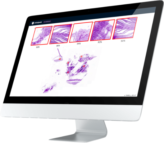
Mindpeak's solution
We solve these challenges by building systems based on artificial intelligence (AI) that work out of the box. Our 0-click solutions are the first ever commercially available products that are able to instantly visualize the detected tumor cells in arbitrary images of IHC-stained breast cancer tissue without the need for manual fine-tuning.

“Mindpeak […] builds extremely good algorithms for the evaluation of immunohistochemical staining using artificial intelligence.”
XTRA, Sysmex Magazin 01/2020
PD Dr. Lukas Heukamp, Head of Molecular Pathology, Haematopathology Hamburg
Find out more about our AI-based diagnostics


.webp)
